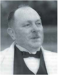
Eponymous Ophthalmologists - Alfred Vogt
EuroTimes looks at the stories behind the names.

Roibeard O’hEineachain
Published: Friday, September 30, 2022
Alfred Vogt 1879–1943
The man behind the palisades.
Alfred Vogt was a Swiss ophthalmologist whose many important contributions to his field of study and practice include the invention of slit lamp microscopy and the description of the ocular structures and pathological signs that bear his name.
Born October 31, 1879, in the village of Menziken, Aargau, Switzerland, Vogt began his study of medicine in 1899 in Zurich before transferring to the University of Basel. There, while under the mentorship of Prof Karl Mellinger, Vogt published his dissertation about “the detrimental effects of artificial aniline dyes on the eye”.i
Receiving his medical doctorate in 1904 and starting a private practice in 1906, Vogt was appointed chief of the Aargau Cantonal Hospital by 1909. His alma mater appointed him professor extraordinarius and director of its eye clinic in 1917.ii
Vogt recognised the potential of the slit lamp in studying the eye’s morphology and pathology early on—combining the device with a microscope in 1913 in a period of discovery for the pioneer. For it was also around this time he found using a redfree light revealed the true yellow colour of the macular lutea.
THE FAMOUS PALISADES
Continuing his series of firsts, Vogt became the first ophthalmologist to perform direct examination of the corneal endothelium in 1918. During the course of this work he identified many anatomical structures and pathological signs that bear his name, including the palisades of Vogt, the radially oriented fibrovascular ridges in the limbus that contain stem cells; Vogt’s striae, fine stress lines within the cornea in eyes with keratoconus; and the limbal girdle of Vogt, a corneal opacity that occurs in an arc concentric pattern and is adjacent to the limbus within the palpebral fissure at 3 and 9 o’clock.
Three years later, he published the first edition of his three-volume textbook on slit lamp microscopy, “Atlas der Spaltlampenmikroskopie des lebenden Auges: mit Anleitung zur Technik und Methodik der Untersuchung” (Atlas of slit lamp microscopy of the living eye: with instructions on the technique and methodology of the examination).
Vogt spent 1920 through 1922 as President of the Swiss Ophthalmological Society. One of his notable acts as president was hosting the first international symposium on slit lamp biomicroscopy in 1922—more than 30 European ophthalmologists (and even one doctor from Japan) attended.
His next appointment was professor ordinarius and director of the University of Zurich’s eye clinic in 1923, where he broadened his investigations into vitreoretinal diseases and began treating retinal detachment by sealing the retinal tear—which later led him to publish a major work on retinal detachment. He also introduced the cyclodiathermy technique for treating glaucoma.
The many awards he received during his life included the Donders Medal of the Dutch Ophthalmological Society in 1932, the Cothenius Medial of the Leopoldina in 1939, the Gonin Medal in 1941, and the Gullstrand Medal of the Swedish Medical Society in 1942.
Sources:
Frederick W Stocker, Arch Ophthalmol. 1944; 31(2): 172–174.
Martin Wenzel. “Why Specular Microscopy?”, Specular Microscopy of Intraocular Lenses: Atlas and Textbook for Slit-Lamp and Specular Microscopic Examinations, 2–4. Stuttgart: Thieme, 1993.
i–ii RD Gerste, Ophthalmologica 2021; 244(1): 1–17.