Virtual Pinhole
New acrylic single-focus biconvex lens for presbyopia.

Roibeard O’hEineachain
Published: Friday, November 1, 2019
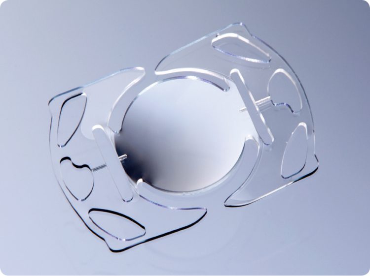 The plate-haptic SC11 intraocular lens. Image courtesy of J Stuart Cumming MD
A new plate-haptic intraocular lens based on a
novel optical approach can provide vision at all distances without dysphotopsias, according to its inventor, J Stuart Cumming MD, Laguna Beach, California, USA.
The lens, the SC11, from CORD LLC. (Cumming Ophthalmic Research & Development), is composed of a hydrophobic acrylic material and has a monofocal biconvex optic and opposing plate haptics, each with two paddle-like proximal extensions designed to partially surround the optic. The plate-haptic/paddle structures are semi-rigid longitudinally but flexible transversely, allowing the SC11 to be folded and implanted through a 2.5mm incision.
“The IOL’s structure with a fixed length, longer than the bag diameter, is designed to consistently place the lens optic far back in the 5mm void left after removing the crystalline lens. This, in turn provides a narrow light cone to increase the depth of focus to provide a virtual pinhole effect,” Dr Cummings told EuroTimes in an interview. The new SC11 lens is modelled after CORD’s SC9 silicone lens, currently in a phase II trial by the US FDA, he added.
In this exploratory trial, investigating both the lens and two insertion devices (Medicel, Switzerland) the method of implantation was finalised. Thirty-eight sequential patients underwent implantation of the lens by Juan Batlle MD, Centro de Microcirugia Ocular Y Laser, Dominican Republic. All patients with cataracts were included. Ten had a preoperative cylinder of 1.0 dioptre or more. Twenty-nine were included in the table. Nine were omitted, four with a BCDVA greater than 20/30, two with surgical complications, one with pre- and post-op cylinders of 2.0D, and two that were uncooperative.
Two different A-constants were used, 119.0 and 120.0, to calculate the lens powers. Lenses were available in 1D steps. Surgery was performed under topical anaesthesia, with a clear corneal incision and phacoemulsification.
Broad range of vision
Of 22 eyes with preoperative K readings of up to 1.08D, including 10 with postoperative refractive cylinders of 1D or more, 13/22 (59%) had uncorrected distance visions of 20/25 or better, and 18/22 (82%) 20/30 or better.
In addition, uncorrected near visual acuity was 20/25 or better in 27%, 20/32 or better in 82% and 20/40 or better in 91%. It would be expected that with good uncorrected distance and near vision, the intermediate visions would be equivalent with a single focus optic. Unexplainably the recorded uncorrected intermediate visual acuity was 20/20 or better in 5%, 20/25 or better 18%, 20/32 or better in 55% and 20/40 or better in 27%.
Furthermore, in 14 eyes, uncorrected distance, intermediate and near visual acuities after cycloplegia were as a good or better than they were before cycloplegia. Moreover, the amount of postoperative cylinder appeared to have no impact on uncorrected visual acuity with or without cycloplegia.
The accommodative effect does not derive from a forward movement of the optic during contraction of the ciliary muscle, which was the proposed mechanism behind the Crystalens, which Dr Cumming developed in the 1990s. Instead the accommodative effect is a result of the greater distance between the posterior surface of the cornea and the front surface of the IOL, which, in turn, results in a narrower cone of light leaving the consistently posterior located optic to reach the retina. This creates a virtual pinhole effect. “The pinhole effect is demonstrated by the similarity of the uncorrected visions with and without cycloplegia,” Dr Cumming said.
He added that his own previous research has shown that the postoperative position of the optic on the anterior-posterior axis is much more consistent with plate-haptic IOLs than with loop-haptic IOLs. That is, the distance between the posterior surface of the cornea and the anterior surface of the optic can vary by as much as 3.15mm with loop-haptic IOLs, compared to a variation of only 1.45mm with the posteriorly locating plate-haptic lenses.
Furthermore, because of their much more posterior position in the capsular bag tamponading the vitreous, plate-haptic IOLs appear to provide some protection against retinal detachment compared to loop-haptic IOLs, Dr Cumming said. In a study in which he and his associates compared outcomes in 1,750 eyes implanted with plate-haptic lenses and 1,857 eyes with loop-haptic lenses. There were 16 retinal detachments, all in the loop-haptics group.
“In loop-haptic IOLs the optic can be located in a forward position, increasing the post-operative volume of the vitreous cavity, allowing the solid vitreous more mobility to tug on the retina and cause a detachment,” he said.
He added that although acrylic IOLs have historically been much more prone to dysphotopsias, no patients reported the phenomenon with the new presbyopic plate-haptic acrylic IOL.
With regard to posterior capsule opacification (PCO), Dr Cumming noted that around 90% of eyes with the new lens require YAG-laser capsulotomy within one year. The opacification’s symptomatic manifestation generally occurs in the form of a reduction in near visual acuity. However, following YAG laser capsulotomies, patients generally recover their near visual acuity, and so far, there have been no cases of retinal detachment and only one case of cystoid macular oedema.
During the study, he and his associates evaluated two Medicel inserter models designed to deliver the lens into the eye with the correct side up. An FDA study with the lens is due to begin soon.
Stuart Cummings: jscumming@gmail.com
The plate-haptic SC11 intraocular lens. Image courtesy of J Stuart Cumming MD
A new plate-haptic intraocular lens based on a
novel optical approach can provide vision at all distances without dysphotopsias, according to its inventor, J Stuart Cumming MD, Laguna Beach, California, USA.
The lens, the SC11, from CORD LLC. (Cumming Ophthalmic Research & Development), is composed of a hydrophobic acrylic material and has a monofocal biconvex optic and opposing plate haptics, each with two paddle-like proximal extensions designed to partially surround the optic. The plate-haptic/paddle structures are semi-rigid longitudinally but flexible transversely, allowing the SC11 to be folded and implanted through a 2.5mm incision.
“The IOL’s structure with a fixed length, longer than the bag diameter, is designed to consistently place the lens optic far back in the 5mm void left after removing the crystalline lens. This, in turn provides a narrow light cone to increase the depth of focus to provide a virtual pinhole effect,” Dr Cummings told EuroTimes in an interview. The new SC11 lens is modelled after CORD’s SC9 silicone lens, currently in a phase II trial by the US FDA, he added.
In this exploratory trial, investigating both the lens and two insertion devices (Medicel, Switzerland) the method of implantation was finalised. Thirty-eight sequential patients underwent implantation of the lens by Juan Batlle MD, Centro de Microcirugia Ocular Y Laser, Dominican Republic. All patients with cataracts were included. Ten had a preoperative cylinder of 1.0 dioptre or more. Twenty-nine were included in the table. Nine were omitted, four with a BCDVA greater than 20/30, two with surgical complications, one with pre- and post-op cylinders of 2.0D, and two that were uncooperative.
Two different A-constants were used, 119.0 and 120.0, to calculate the lens powers. Lenses were available in 1D steps. Surgery was performed under topical anaesthesia, with a clear corneal incision and phacoemulsification.
Broad range of vision
Of 22 eyes with preoperative K readings of up to 1.08D, including 10 with postoperative refractive cylinders of 1D or more, 13/22 (59%) had uncorrected distance visions of 20/25 or better, and 18/22 (82%) 20/30 or better.
In addition, uncorrected near visual acuity was 20/25 or better in 27%, 20/32 or better in 82% and 20/40 or better in 91%. It would be expected that with good uncorrected distance and near vision, the intermediate visions would be equivalent with a single focus optic. Unexplainably the recorded uncorrected intermediate visual acuity was 20/20 or better in 5%, 20/25 or better 18%, 20/32 or better in 55% and 20/40 or better in 27%.
Furthermore, in 14 eyes, uncorrected distance, intermediate and near visual acuities after cycloplegia were as a good or better than they were before cycloplegia. Moreover, the amount of postoperative cylinder appeared to have no impact on uncorrected visual acuity with or without cycloplegia.
The accommodative effect does not derive from a forward movement of the optic during contraction of the ciliary muscle, which was the proposed mechanism behind the Crystalens, which Dr Cumming developed in the 1990s. Instead the accommodative effect is a result of the greater distance between the posterior surface of the cornea and the front surface of the IOL, which, in turn, results in a narrower cone of light leaving the consistently posterior located optic to reach the retina. This creates a virtual pinhole effect. “The pinhole effect is demonstrated by the similarity of the uncorrected visions with and without cycloplegia,” Dr Cumming said.
He added that his own previous research has shown that the postoperative position of the optic on the anterior-posterior axis is much more consistent with plate-haptic IOLs than with loop-haptic IOLs. That is, the distance between the posterior surface of the cornea and the anterior surface of the optic can vary by as much as 3.15mm with loop-haptic IOLs, compared to a variation of only 1.45mm with the posteriorly locating plate-haptic lenses.
Furthermore, because of their much more posterior position in the capsular bag tamponading the vitreous, plate-haptic IOLs appear to provide some protection against retinal detachment compared to loop-haptic IOLs, Dr Cumming said. In a study in which he and his associates compared outcomes in 1,750 eyes implanted with plate-haptic lenses and 1,857 eyes with loop-haptic lenses. There were 16 retinal detachments, all in the loop-haptics group.
“In loop-haptic IOLs the optic can be located in a forward position, increasing the post-operative volume of the vitreous cavity, allowing the solid vitreous more mobility to tug on the retina and cause a detachment,” he said.
He added that although acrylic IOLs have historically been much more prone to dysphotopsias, no patients reported the phenomenon with the new presbyopic plate-haptic acrylic IOL.
With regard to posterior capsule opacification (PCO), Dr Cumming noted that around 90% of eyes with the new lens require YAG-laser capsulotomy within one year. The opacification’s symptomatic manifestation generally occurs in the form of a reduction in near visual acuity. However, following YAG laser capsulotomies, patients generally recover their near visual acuity, and so far, there have been no cases of retinal detachment and only one case of cystoid macular oedema.
During the study, he and his associates evaluated two Medicel inserter models designed to deliver the lens into the eye with the correct side up. An FDA study with the lens is due to begin soon.
Stuart Cummings: jscumming@gmail.com