Cheryl Guttman Krader reports from ASCRS 2021 in Las Vegas, USA.
Sight-threatening complications, including blindness, are being reported with unregulated anterior segment devices designed to change the cosmetic appearance of healthy eyes. The moral of the story, therefore, is simple and clear—
“Do not insert unregulated devices in or around the eye,” said Sonal S Tuli MD, MEd.
Dr Tuli reviewed complications associated with cosmetic iris implants, eye jewellery, eyeball tattooing, keratopigmentation, and coloured cosmetic contact lenses.
She differentiated the cosmetic iris implants from the devices approved by the US Food and Drug Administration and other regulatory agencies for patients with aniridia. Unlike the latter devices put in the capsular bag after cataract removal, the cosmetic iris implants are placed in the anterior chamber over the iris to change iris colour in persons who otherwise have healthy eyes.
The cosmetic iris implants have irregularities on their posterior surface and small prongs that insert into the anterior chamber angle. While these design features presumably serve to help maintain positional stability of the device, they cause iris and trabecular meshwork damage from chafing that can result in the development of the uveitis-glaucoma-hyphaema (UGH) syndrome.
Other complications associated with these devices include cataract, cystoid macular oedema, corneal oedema, corneal neovascularisation, and keratic precipitates. Even after undergoing device explantation, patients have suffered permanent visual loss and persistent severe glaucoma requiring surgical management, Dr Tuli said.
Eye jewellery inserted as an adornment under the conjunctiva has also come into vogue in recent years. Because these articles are typically platinum, which is inert, they do not tend to cause inflammation.
“Thus far, no significant complications have been reported with the use of eye jewellery. Nevertheless, the potential for problems remains due to the theoretic risks for jewellery migration, infection, inflammation, and perforation,” Dr Tuli said.
Eyeball tattooing, a procedure done for cosmetic reasons in which dye is injected under the conjunctiva, has also been associated with serious complications.
“Some individuals undergo eyeball tattooing because they want to permanently change their eye colour, whereas others (erroneously) expect it is a way to temporarily change eye colour,” Dr Tuli explained.
The dye used for eyeball tattooing is often the same material used for skin tattooing, which is highly inflammatory in the eye. Furthermore, the tattooing is usually done without microscopic visualisation, proper surgical instruments, or sterile conditions. Therefore, the risks of infection, inflammation with granuloma development, and globe penetration are high.
Highlighting the complications associated with eyeball tattooing, Dr Tuli presented a case reported in the literature in which the dye migrated posteriorly, leading to posterior scleritis and loss of vision. Although treatment with tarsorrhaphy, high-dose corticosteroids, and antibiotics resulted in improvement, the patient was left with a green eye and green eyelids.
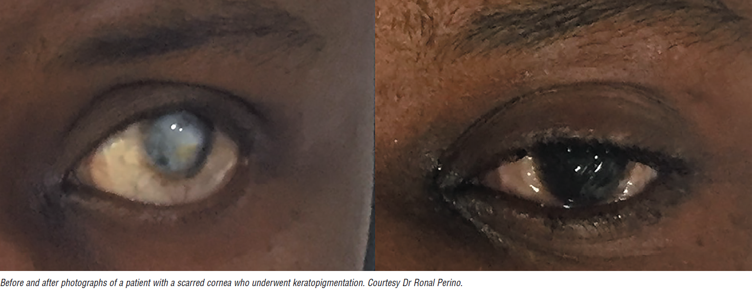
Keratopigmentation is a technique that has been used for centuries to mask disfiguring corneal scars. Today, cornea specialists still do it to improve the cosmetic appearance of blind eyes or as a therapeutic intervention to address functional visual symptoms such as glare from large iris defects. However, untrained persons also perform it, using unregulated materials as a purely cosmetic procedure to change eye colour in healthy eyes.
Historically, keratopigmentation compounds were composed of India ink, soot, or platinum chloride, but recent procedures use micronized mineral pigments with lower inflammatory potential. The pigments may be placed using a needle tattooing technique or in a femtosecond laser-assisted procedure where the laser creates channels in the cornea into which the specialist injects pigments.
Despite the better safety profile of the current generation of mineral pigments used for keratopigmentation, significant complications have been reported, including corneal pannus, inflammation, and pigment migration to other parts of the eye.
“In some cases, the inflammation has been severe enough that it resulted in cornea melting,” Dr Tuli said.
Depending on the pigment material used, interaction with MRI is also a potential concern, as demonstrated by the case of a patient who had undergone keratopigmentation and developed severe eye pain during head MRI.
“Even though micronised mineral pigments are thought to be somewhat ‘MRI compatible’, I warn patients undergoing keratopigmentation for a medical indication about the potential for an MRI interaction, even if the area of tattooing is very small,” Dr Tuli said.
COSMETIC CONTACTS NOT LOOKING GOOD
Cosmetic contact lenses are often used by people dressing up in costume for Halloween and are frequently bought online or at a hair or cosmetic salon. Often unsterile, sometimes previously worn by multiple people, and coming without instructions on cleaning, these non-prescription contact lenses are notorious for causing ocular complications such as corneal abrasions and ulcers, which can result in permanent loss of vision.


