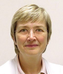
Carina Koppen MD, PhD
Incorporating the current guidelines from the Tear Film Ocular Surface Society’s 2017 dry eye workshop report (TFOS DEWS II) for dry eye disease (DED) diagnosis into one’s daily surgical practice does not require special expertise in ocular surface disease or the purchase of new expensive equipment, said Carina Koppen MD, PhD, University of Antwerp, Belgium.
“Actually, the scheme includes a lot of tests that you are already familiar with, and with a bit of organisation it’s very possible to include these tests in the routine of your daily workflow,” Dr Koppen told the 25th ESCRS Winter Meeting.
The guidelines suggest starting with a series of triaging questions to help rule in dry eye and rule out other ocular surface diseases. They include questions regarding not only the typical DED symptoms of discomfort and their triggering factors, but also questions regarding visual acuity and fluctuating vision. Also important is a risk factor analysis, to obtain a fuller picture of the patient’s overall ocular and systemic health, with questions regarding smoking, medications and contact lens wear.
Dr Koppen noted that the initial parts of the investigation can be greatly facilitated by the use of questionnaires, such as the Ocular Surface Disease Index and SANDE questionnaires. They have the advantage that patients can fill them out in the waiting room, reducing chair time.
SLIT-LAMP EXAMINATION
The next step in the TFOS DEWS II scheme is a systematic slit-lamp examination of the ocular surface, including the eyelids, lashes and margins and the lower and upper conjunctiva. Features to look for include conjunctival folds suggestive of dry eye disease and a frothy aspect of the tears such as occur in the presence of meibomian gland disease.
“You might miss cicatrising conjunctivitis or superior limbic keratoconjunctivitis if you don’t examine the eye in the upward and downward gaze, and epithelial dystrophy, which is actually not so rare if you look for it,” she said.
Following this examination, the clinician needs to determine tear break-up time (TBUT) by instilling a drop of fluorescein. A TBUT of less than 10 seconds is suggestive of tear film instability, a TBUT of less than five seconds is the signature of definitive DED. The residual fluorescein stains areas of epithelial damage and erosions.
“As clinicians we should not forget to systematically examine the eyelid margins and the orifices of the meibomian gland and to squeeze the glands to test their functionality,” she added.
MEASUREMENT OF TEAR OSMOLARITY
Osmolarity is another parameter included in the TFOS DEWS II guidelines. Dr Koppen noted that hyperosmolarity is a hallmark sign of DED. Maximum osmolarity greater than 308mOsmol/L and/ or inter-eye difference of >8mOsmol/L suggest DED. But whether ophthalmic clinicians require an osmolarity metre next to the slit lamp remains a matter of debate. The tests have high specificity (78-to-99%) but a more variable sensitivity (48-to-95%).
“The main problem is that values of osmolarity fluctuate, so it is advised to at least test both eyes. The test would probably be more sensitive if you do several tests per eye but each chip costs money so the test becomes prohibitively expensive if you’re going to test both eyes three times,” Dr Koppen said.
Once it has been ascertained on the basis of these tests that a patient has dry eye disease, the next step is to classify the condition as evaporative or aqueous deficient dry eye. Changes in the tear film lipid layer and the meibomian glands are suggestive of evaporative dry eye, while reductions in the tear meniscus height indicate aqueous deficient dry eye, Dr Koppen said.
“Actually, it is not an either/or situation. Once the patient gets trapped in a vicious circle of dry eye disease then both types will be present in the same patient at a certain point in time,” she added.
Dr Koppen added that multi-functional devices are becoming available for the automation of DED diagnosis with considerable accuracy. They include the Oculus keratograph, a tomographer that also has an inbuilt infrared camera for meibography. It can also perform interferometry of the lipid layer, non-invasive tear film break-up time, tear film meniscus height measurement and lipid layer evaluation. Other dedicated and multifunctional devices have recently been introduced for clinical use.
“If you want to improve your dry eye disease diagnostics, systematically screen for signs and symptoms, listen to the patient’s description of their symptoms of discomfort and fluctuating vision, look at the slit lamp and don’t forget to use a drop of fluorescein for TBUT and staining, which can tell you so much, and don’t forget to inspect and squeeze the meibomian glands,” Dr Koppen concluded.

 Carina Koppen MD, PhD