Angiographic studies and optical coherence tomography (OCT) have found structural evidence suggesting lymphatics drain aqueous humour from subconjunctival blebs used to lower intraocular pressure in glaucoma surgery and treat various eye diseases via drug delivery, Alex Huang MD, PhD reported at the Association for Research in Vision and Ophthalmology 2021 Annual Meeting.
In a study of 10 patients, angiographic imaging of subconjunctival blebs revealed sausage-shaped outflow pathways previously thought to represent lymphatics. Simultaneous OCT imaging showed semilunar valves or flaps that are a lymphatic hallmark, said Dr Huang, Associate Professor at the Doheny Eye Institute and the Stein Eye Institute at the University of California–Los Angeles, USA.
This study provided multimodal outflow/structural imaging evidence confirming the hypothesis that lymphatics drain blebs in live humans, Dr Huang said. Potential implications include the ability to regulate bleb outflow by encouraging or discouraging outflow using drugs affecting the lymphatic system, he added.
“If we can understand this better, we could improve the treatment of eye diseases.”
ANIMAL AND HUMAN STUDIES
Subconjunctival blebs are intentionally created in traditional glaucoma drainage surgery as well as minimally invasive glaucoma surgery (MIGS) devices such as the XEN Gel Stent (Allergan) and the PreserFlo MicroShunt (Santen). Additionally, they develop during subconjunctival drug delivery. Blebs may also occur spontaneously as chemosis due to fluid overload, infection, inflammation, or head and neck surgery, Dr Huang noted.
So how does fluid exit blebs? Mechanisms suggested include leaking into the orbit or through blood vessels. But observation of patients undergoing subconjunctival haemorrhage and dye injections in studies show dispersion through irregular pathways with alternating wide and narrow points resembling sausage links, suggesting the lymphatic system is involved, Dr Huang said.
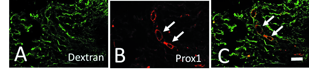
In animal studies using two-photon microscopy and OCT from colleague Goichi Akiyama MD, PhD, Dr Huang found bicuspid valves in the direction of subconjunctival outflow, providing additional evidence of lymphatic involvement. Using molecular markers, they were also able to confirm bleb-related outflow pathways expressed specific lymphatic but not blood vessel markers. (Fig 1)
In his human study, Drs Huang and Jong Young Lee MD injected indocyanine green and subconjunctival lidocaine in macular degeneration patients preparing for intravitreal injection. This enabled ocular surface lymphangiography, which revealed outflow pathways in seven patients. Simultaneous OCT showed the variable lumen diameter and the presence of valve-like structures along outflow pathways. In comparison, aqueous angiography imaging of trabecular outflow (which visualizes veins, not lymphatics) demonstrated even-calibered pathways without valves that took typical venous Y-shaped branching.
Understanding the mechanism is important because pharmacological manipulation of lymphatics could make glaucoma surgery more successful by increasing outflow—or make subconjunctival drug delivery more effective by reducing outflow, Dr Huang concluded.

Alex Huang: ahuang@doheny.org or alhuanga@yahoo.com