Myopic foveoschisis

Early intervention correlates with better functional outcome

Myopic foveoschisis
Cheryl Guttman Krader
Published: Friday, March 1, 2019
[caption id="attachment_14161" align="alignleft" width="945"] 
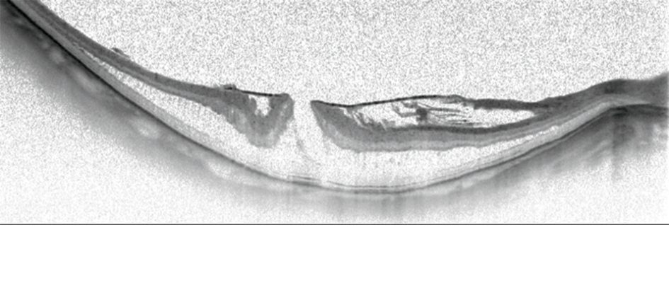
Horizontal OCT scan of a myopic foveoschisis with a premacular membrane[/caption] Preoperative visual acuity is the main factor influencing functional outcomes after vitrectomy for myopic foveoschisis. With this information in mind, retina specialists aiming to improve their results should consider operating sooner rather than later, said Ramin Tadayoni MD, PhD, at the 18th EURETINA Congress in Vienna, Austria. “Of course, we can also do better for our patients by using modern surgical techniques and imaging technologies. But, we will have better results if the indication for surgery is good. If the goal is to achieve BCVA [best-corrected visual acuity] better than 20/40, then surgery should be done when the vision is 20/50 or better. We should not wait for the vision to get worse,” said Dr Tadayoni, Professor of Ophthalmology, Paris 7 University, Lariboisière Hospital & OphtalmoPôle, Paris, France.
Ramin Tadayoni MD, PhD
Dr Tadayoni and colleagues conducted a retrospective study to identify preoperative factors influencing visual recovery after pars plana vitrectomy for myopic foveoschisis. The study, which has been published (Lehmann M, et al. Retina. 2017 Dec 1. Epub ahead of print), included 66 eyes of 65 consecutive highly myopic patients with a mean postoperative follow-up of 14 months. The eyes were stratified into quartiles according to preoperative BCVA (≥20/50, 20/60 to 20/80, 20/100 to 20/125, and ≤20/160) and into three groups based on their preoperative foveal status (simple foveoschisis, foveoschisis with foveal detachment, foveoschisis with macular hole). All four BCVA subgroups benefited with a significant improvement in BCVA, and the worse the baseline vision, the greater the gain. However, mean BCVA for patients in the lower three quartiles did not reach the level achieved by the subgroup with BCVA ≥20/50, and only the highest quartile achieved a mean final BCVA better than 20/40. Predictors for final BCVA were assessed using an analysis of covariance performed with the anatomical status at baseline adjusted for baseline BCVA, spherical equivalent, axial length, central foveal thickness, and age. Of all of the preoperative variables, BCVA was the only factor that correlated with postoperative vision. Foveal status was associated with vision outcome in univariate analysis, but it was not an independent predictor of final BCVA after adjusting for baseline vision, Dr Tadayoni reported. “The literature suggests that vision gains are better in eyes with foveal detachment than in those with foveoschisis. The explanation is probably that many eyes with foveal detachment present with low vision and therefore have greater gains. But vision in an eye with foveoschisis and low vision evolves exactly like in an eye with a foveal detachment and low vision,” he said. Practically none of the eyes operated on with poorer vision achieved final BCVA of 20/40 or better. Seven (10.6%) eyes lost vision from baseline to last visit because of macular changes. “Interestingly, most of the complications were concentrated in the low vision group. It seems if we wait too long because we are being cautious, we are going to more risky surgery later,” Dr Tadayoni said. EXTRAPOLATING EVIDENCE TO CLINICAL CARE Dr Tadayoni acknowledged that the findings of this retrospective study do not provide definitive guidance for surgical decisions, but in the absence of evidence from a randomised interventional study comparing early versus late surgery, they provide useful information. “This is a simple piece of information that I think retina specialists can integrate into their practice, but it should not be interpreted as suggesting that all patients should be operated at the stage when vision is better than 20/50,” he said. “In practice, we need to take into account the preoperative status of the eye, whether there is a foveal detachment, if there is a high risk of having a macular hole and also the patient’s preferences and needs. For patients who have 20/50 vision and are happy, we can wait. “These patients, however, need to be informed that if they wait until their vision worsens, they may recover vision, but it may not reach 20/40.”
Tags: myopic foveoschisis, vitrectomy
Latest Articles
Simulators Benefit Surgeons and Patients

Helping young surgeons build confidence and expertise.

Read more...

How Many Surgeries Equal Surgical Proficiency?

Internet, labs, simulators, and assisting surgery all contribute.

Read more...

Improving Clinical Management for nAMD and DME

Global survey data identify barriers and opportunities.

Read more...

Are Postoperative Topical Antibiotic Drops Still Needed?

Cataract surgeons debate the benefits of intracameral cefuroxime prophylaxis.

Read more...

Emerging Technology for Detecting Subclinical Keratoconus

Brillouin microscopy shows promise in clinical studies.

Read more...

Knowing Iris Repair: Modified Trifold Technique

Part eight of our series covers the modified trifold technique for large iris defects.

Read more...

It’s All About Biomechanics!

Increasing the pool of patients eligible for refractive surgery.

Read more...

Uncovering More Safe and Quick Options

Different strategies, such as PresbyLASIK, can offer presbyopes good outcomes.

Read more...

Topography-Guided PRK for Keratoconus

Improving visual acuity in patients with keratoconus.

Read more...

Defining AMD Treatment Protocol

Treatments trending to fewer injections for better results.

Read more...