Endopthalmitis and keratoplasty

Rise in fungal endophthalmitis reflects changing keratoplasty practice

Endopthalmitis and keratoplasty
Dermot McGrath
Dermot McGrath
Published: Friday, February 1, 2019
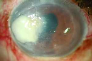
Fungal infiltrate following DSAEK
The trend in corneal transplantation towards endothelial keratoplasty (EK) procedures at 
the expense of penetrating keratoplasty may be partially responsible for an increase in cases of fungal endophthalmitis in recent years, according to Sathish Srinivasan FRCOphth. “Although fungal infections can occur with all types of transplant, whether penetrating keratoplasty (PK) or lamellar transplants, the evidence in the scientific literature indicates that endophthalmitis after EK is primarily fungal in nature whereas infection after PK is mostly bacterial,” Dr Srinivasan told delegates attending the 9th EuCornea Congress in Vienna. Analysing 354,930 transplants performed in the United States from 2007 to 2014, the Eye Bank Association of America (EBAA) found primary graft failure the most common adverse reaction (319 cases or 0.09%) followed by endophthalmitis in 99 cases or 0.028%. The procedure type predominantly associated with endophthalmitis was endothelial keratoplasty in 61%, followed by PK in 37% and keratoprosthesis in 1%. Of the 77 culture-positive cases in the EBAA report, the majority were caused by fungal infection in 65% versus bacterial infection in 35%. The high association between fungal infection and EK and tissue prepared in the eye bank merits further study, said Dr Srinivasan, Consultant Corneal Surgeon at University Hospital Ayr, Ayr, Scotland.
Fungal infiltrate following PK
“We need more data from Europe and elsewhere to confirm the data from the US on the increased risk of using pre-cut tissues from eye banks compared to surgeon-prepared tissue after EK. Several factors such as the corneal storage medium, increased use of antibiotics and increased warming period time to prepare tissue have all been suggested as possible reasons for the increased incidence of fungal causative pathogens,” he said. Expanding on these factors, Dr Srinivasan said that there is currently no routine use of an antifungal agent to supplement corneal storage medium in the United States, while the increased use of broad-spectrum antibiotics before and after surgery creates a non-competitive environment that allows fungi to thrive. Finally, the increased warming period time associated with preparing EK tissue in the eye bank is thought to favour increased fungal proliferation, he added. The scientific literature on endophthalmitis after keratoplasty is quite threadbare, noted Dr Srinivasan, with only a handful of papers putting the spotlight on one of the most feared complications of ocular surgery. “The majority of studies deal with endophthalmitis after PK, so there is a clear need for better data to reflect changing trends in corneal transplantation. Unlike cataract surgery, where there is only one single intervention, the causes of endophthalmitis following keratoplasty are multifactorial and can be related to donor, recipient and storage media factors, which vary greatly in different parts of the world,” he said. Looking at the incidence of endophthalmitis after keratoplasty, a UK study of 11,320 transplant recipients between 1999 and 2006 reported an endophthalmitis incidence of 0.67% after primary PK. The incidence of endophthalmitis occurring within six weeks of surgery was 0.16% and graft survival after endophthalmitis was 27% at five years, noted Dr Srinivasan. “I think this is one of the key take-home messages – if the surgery was a PK and the patient develops bacterial endophthalmitis then the final survival rate is dramatically reduced. Of the 76 cases in the UK study the predominant bugs found were pseudomonas, staphylococcus and streptococci in about 69% of cases,” he said. Factors associated with endophthalmitis were donor cause of death (infection), high-risk cases and indication for 
corneal transplantation. “What stood out was that all the patients who had interventions for therapeutic keratoplasty had a slightly higher risk of developing postoperative endophthalmitis. It was also interesting to note that if the donor’s cause of death was infection such as septicaemia then the donor's corneas were at higher risk of developing infection,” he said. Summing up, Dr Srinivasan said that endophthalmitis remains a serious issue after corneal graft surgery, with those affected having reduced graft survival and poor visual outcomes. “Management of the identified recipient and donor risk factors are important to reduce endophthalmitis risk, so we definitely need more data to shed light 
on these factors in the future,” he said.
Tags: cornea, endophthalmitis
Latest Articles
Simulators Benefit Surgeons and Patients

Helping young surgeons build confidence and expertise.

Read more...

How Many Surgeries Equal Surgical Proficiency?

Internet, labs, simulators, and assisting surgery all contribute.

Read more...

Improving Clinical Management for nAMD and DME

Global survey data identify barriers and opportunities.

Read more...

Are Postoperative Topical Antibiotic Drops Still Needed?

Cataract surgeons debate the benefits of intracameral cefuroxime prophylaxis.

Read more...

Emerging Technology for Detecting Subclinical Keratoconus

Brillouin microscopy shows promise in clinical studies.

Read more...

Knowing Iris Repair: Modified Trifold Technique

Part eight of our series covers the modified trifold technique for large iris defects.

Read more...

It’s All About Biomechanics!

Increasing the pool of patients eligible for refractive surgery.

Read more...

Uncovering More Safe and Quick Options

Different strategies, such as PresbyLASIK, can offer presbyopes good outcomes.

Read more...

Topography-Guided PRK for Keratoconus

Improving visual acuity in patients with keratoconus.

Read more...

Defining AMD Treatment Protocol

Treatments trending to fewer injections for better results.

Read more...