Diabetic retinopathy today
Paradigm shift in understanding and treatment of DR.

Dermot McGrath
Published: Saturday, February 1, 2020
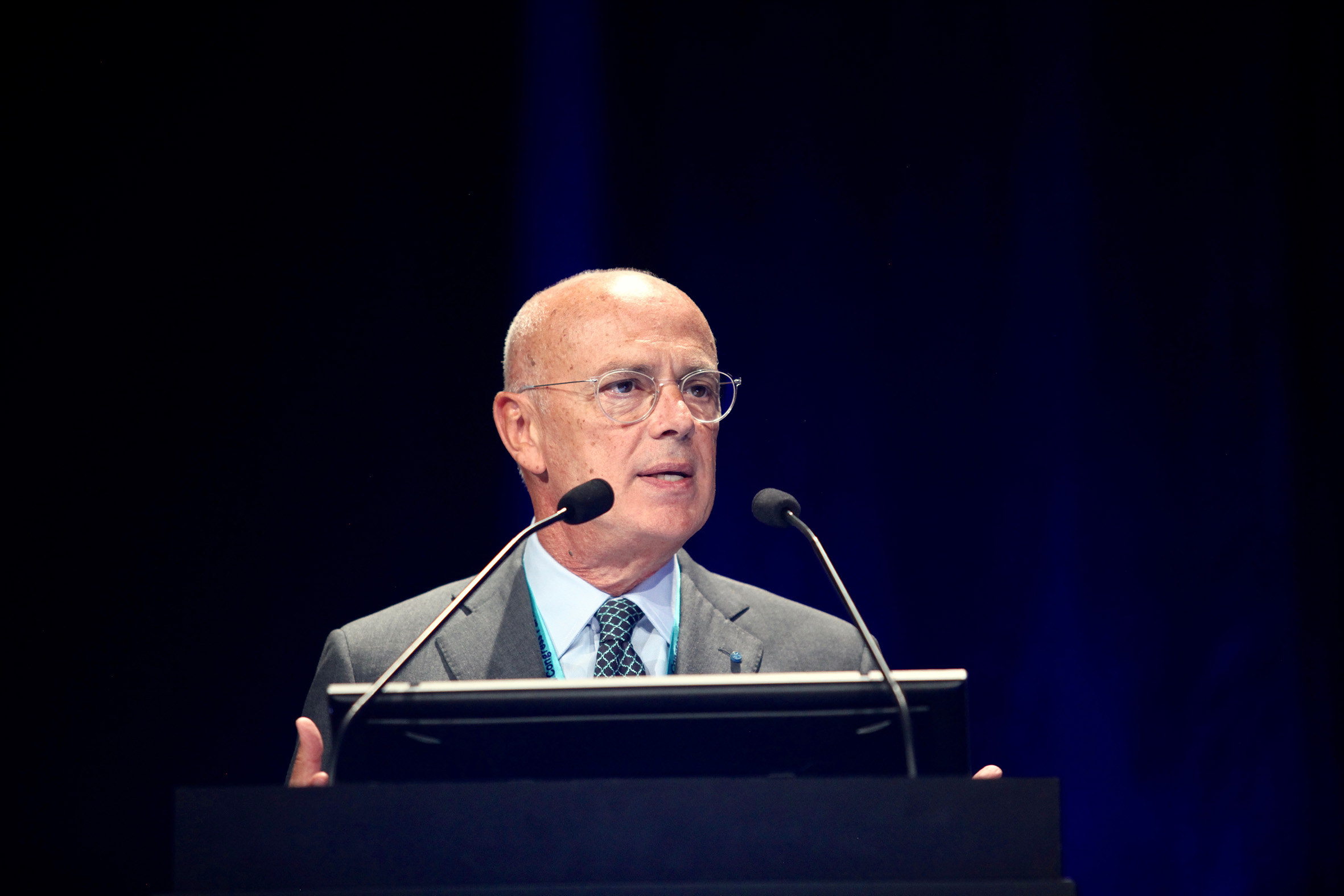
 Professor Francesco Bandello MD, FEBO.
Recent decades have seen a profound transformation in the understanding of the complex pathophysiology of diabetic retinopathy (DR), with the evolution of new treatment strategies that move beyond purely metabolic control to try to mitigate the sight-threatening ocular complications of the disease, according to Professor Francesco Bandello MD, FEBO.
“We have witnessed a paradigm shift in the interpretation of diabetic retinopathy in the last 50 years from a disease of pure microangiopathy to one of neurovascular coupling dysfunction in which all the microvascular districts such as the iris, optic nerve, and macula are involved,” said Prof Bandello, in his EURETINA Medal Lecture at the 19th EURETINA Congress in Paris.
In a broad overview of the evolution of knowledge pertaining to DR, Prof Bandello said its development reminded him of the way historians divide the study of their discipline into two broad categories: prehistory, before the introduction of written records, and history.
“This is pretty much the same with DR. The prehistory period was when the ophthalmoscope was not available, and theories were proposed on the basis of intuition but without the opportunity to witness directly what they were talking about. This changed after the introduction of ophthalmoscopy in the mid-19th Century,” said Prof Bandello, Full Professor and Chairman at the Department of Ophthalmology, University Vita-Salute, Scientific Institute San Raffaele, in Milan, Italy.
The very early history of DR research was hampered to a significant degree by the celebrated ophthalmologist Albrecht von Graefe’s claim in 1856 that there was no proof of a causal relationship between diabetes and retinal complications, noted Prof Bandello.
“Although von Graefe got it wrong, there was another giant of ophthalmology at the time, Eduard Jaeger, who said the opposite and was able to recognise lesions that were due to diabetes,” he said.
Breakthroughs
The introduction of fluorescein angiography (FA) and laser photocoagulation proved to be a game-changer in the modern era of DR diagnosis and treatment, said Prof Bandello.
“The combination of these two events, one for diagnosis and one for therapy, made a huge difference in understanding the pathogenesis of retinal lesions in diabetes and to begin to treat them with laser,” he said.
In the 1960s, key fluorescein studies of the retina in diabetics were carried out by Scott and Dollery, along with the pioneering work of John Gass, who first described retinal ischaemia and the appearance of new vessels with hyperfluorescent aspects using FA. Prof Bandello also cited the work of Koichi Shimizu in describing mid-peripheral fundus involvement in DR and also the relationship between ischaemia and new vessels.
Another important milestone came with the publication of the results of two key studies: the Diabetic Retinopathy Study, which showed that laser photocoagulation reduced the two-year incidence of severe visual loss by more than half in eyes with proliferative diabetic retinopathy, and the Early Treatment Diabetic Retinopathy Study (ETDRS), which demonstrated that focal macular laser reduced the risk of moderate vision loss by up to 50% in eyes with clinically significant macular oedema.
Other major advances came with the introduction of optical coherence tomography (OCT) into clinical practice followed closely by intravitreal steroids and anti-VEGF therapies.
“I think we would all agree that both of these developments completely transformed the way we diagnose, treat and follow-up our patients,” said Prof Bandello.
Prof Bandello cited the work of his own research team over the years in elucidating the complex pathophysiology of DR.
“We were among the first to underline the importance of FA to understand the pathogenetic mechanism between the blood-retinal-barrier (BRB) breakdown and the appearance of macular oedema. We have also worked hard to promote the concept of lighter-intensity laser modalities for optimal therapeutic effect in patients with clinically significant macular oedema,” he said.
New classification system
With so many treatment options currently available, Prof Bandello said that a new classification system of DME into distinct categories will help orient treatment choice.
“I like the concept of subdividing DME into different sub-groups because only then will be able to select the best treatment for each individual patient. We cannot proceed as before using only one therapeutic option, which was laser. We have developed an algorithm to help guide the treatment once the subtype has been identified,” he said.
Prof Bandello added that the integration of standard investigations with new diagnostic techniques will allow prompt recognition and personalised treatment of both retinopathy and maculopathy in the near future.
The need for such an evolution is all the more evident given the impending global epidemic of diabetes and its impact on health systems everywhere, warned Prof Bandello.
“This is important because we know diabetes is exploding worldwide and as ophthalmologists we must do what we can to prepare for the consequences of that for our patients’ vision. We need to be able to work with our public health administrators and politicians in order to apply the best of what we have today for the diagnosis and treatment of DR,” he concluded.
Francesco Bandello:
bandello.francesco@hsr.it
Tags: diabetic retinopathy
Latest Articles
Towards a Unified IOL Classification
The new IOL functional classification needs a strong and unified effort from surgeons, societies, and industry.
The 5 Ws of Post-Presbyopic IOL Enhancement
Fine-tuning refractive outcomes to meet patient expectations.
AI Shows Promise for Meibography Grading
Study demonstrates accuracy in detecting abnormalities and subtle changes in meibomian glands.
Are There Differences Between Male and Female Eyes?
TOGA Session panel underlined the need for more studies on gender differences.
Simulating Laser Vision Correction Outcomes
Individualised planning models could reduce ectasia risk and improve outcomes.
Need to Know: Aberrations, Aberrometry, and Aberropia
Understanding the nomenclature and techniques.
When Is It Time to Remove a Phakic IOL?
Close monitoring of endothelial cell loss in phakic IOL patients and timely explantation may avoid surgical complications.
Delivering Uncompromising Cataract Care
Expert panel considers tips and tricks for cataracts and compromised corneas.
Organising for Success
Professional and personal goals drive practice ownership and operational choices.