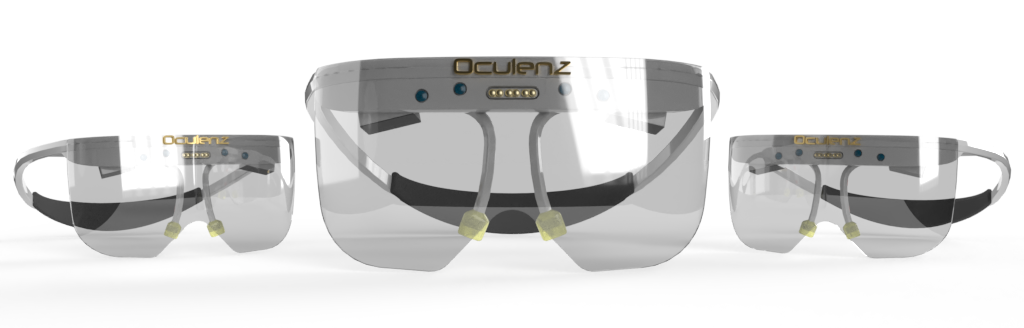
Augmented Reality: New tool in ophthalmology
New device provides visual rehabilitation for AMD patients

Leigh Spielberg
Published: Monday, February 1, 2021
 Linda Lam, MD, MBA
New device provides visual rehabilitation for AMD patients
Published: Monday, February 1, 2021
Linda Lam, MD, MBA