Artificial intelligence and retinal disease
AI is helping to level playing field in DR management

Dermot McGrath
Published: Friday, November 1, 2019
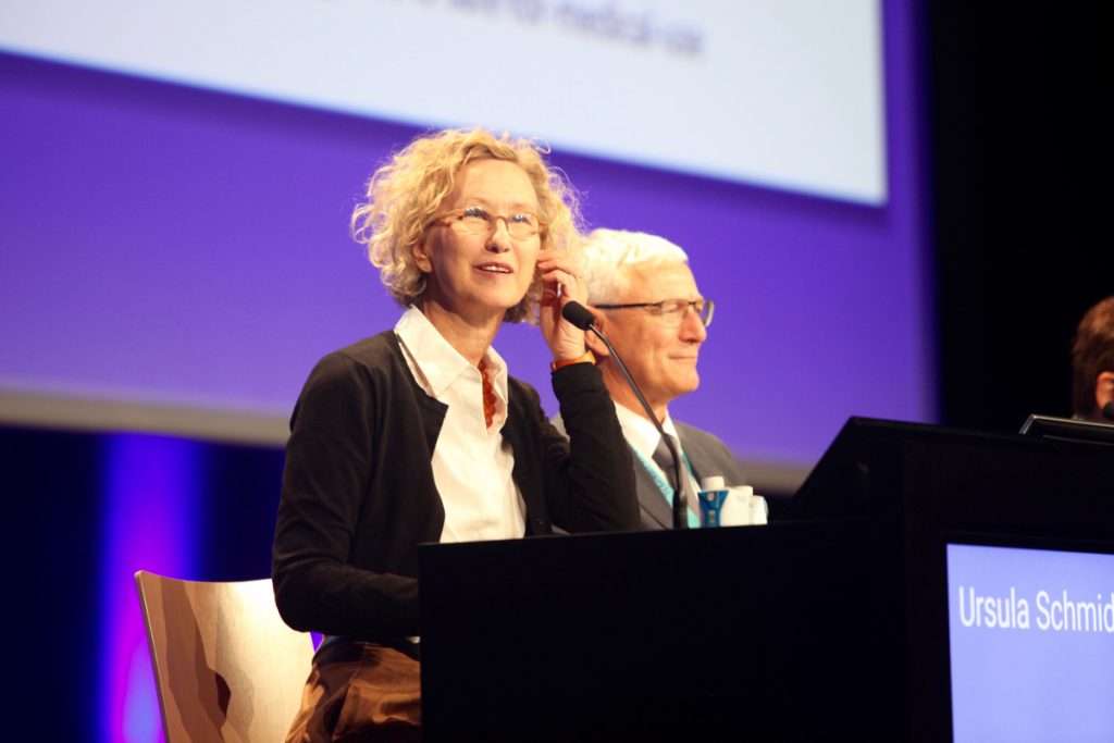 Ursula Schmidt-Erfurth MD, PhD, at the 19th EURETINA Congress in Paris, France[/caption]
Artificial intelligence (AI) is already making a direct impact in the screening and management of diabetic eye disease in the face of a growing epidemic of diabetes worldwide, Ursula Schmidt-Erfurth MD, PhD, said at the 19th EURETINA Congress in Paris.
“AI-based image analysis offers a rapid, cost-effective, precise and reliable screening and monitoring of diabetic retinopathy (DR) and diabetic macular oedema (DME). This technology is not here to replace doctors but to assist them in managing their diabetic patients more efficiently,” she said.
Prof Schmidt-Erfurth told delegates that machine learning and AI applications will become increasingly important in tackling the impending global epidemic of diabetes-related ocular disease.
“Diabetes mellitus affects 420 million people who will eventually develop diabetic retinopathy, which includes over 20 million people with macular disease. On the other hand, there are only 210,000 retinologists working in the world to try to keep this under control. This clear disproportion highlights the need for AI support to manage such a widespread disease,” she said.
The first USA Food and Drug Administration (FDA) clearance for an autonomous AI-diagnostic system (IDx-DR) to automatically detect more than mild DR in previously undiagnosed adults with diabetes was granted in 2018, explained Prof Schmidt-Erfurth.
“This is a screening device and not a monitoring device. It does not replace eye doctors but it brings the right patients to the eye doctors at the right time. The FDA decision was based on proof of principle studies, which showed that AI is as accurate or better than the human eye in the analysis of digital fundus images,” she said.
The AI system also integrates quality assessment of the underlying image acquisition, which is critical in providing a clear diagnostic conclusion and a clinically plausible diagnostic decision, noted Prof Schmidt-Erfurth.
Approval of the IDx-DR system followed a study in 900 diabetic patients at 10 primary care centres in the United States, with the algorithm trained to detect more than mild DR and/or DME versus mild or no DR.
“It is not about detecting a single aneurysm at 280 degrees – this device is made to recognise clinically relevant disease,” said Prof Schmidt-Erfurth.
The reference standard was provided by a certified expert reading centre, which was offered four ultra-wide-field stereo images for determination of ETDRS scores and also optical coherence tomography (OCT) for DME diagnosis by three independent readers.
“This was unfair competition because the human experts were much better equipped. Despite this, the AI system provided a sensitivity of 87% and a specificity of 91%, outperforming the superiority endpoints set by the FDA,” she said.
The initial trial results were subsequently confirmed by a global peer-reviewed validation, which led to an expanded use of AI for diabetic screening in multiple countries in Europe.
AI also has rich potential in the monitoring of DME, which is particularly useful given that most patients who attend clinics are already at an advanced stage of disease and often present with visual loss due to macular oedema, noted Prof Schmidt-Erfurth.
Histology studies have shown that intraretinal fluid typically presents in a cystoid formation, which intensively damages neurosensory structures.
“The more cysts, the greater the vision loss, and the bigger the cysts the more irreversible the vision loss,” she said.
Non-invasive high-resolution OCT imaging not only visualises intraretinal and subretinal fluid but also offers precise measurement of fluid volumes that can then be analysed using a machine-learning algorithm.
“In diabetic microvascular disease, there are a lot of cystic changes in the retina, which clinicians are unable to count and monitor. However, this kind of information is needed if we want to precisely follow and judge disease progression or a good therapeutic response,” she said.
Some recent studies have shown that intraretinal cystoid fluid (IRC) in the central fovea is what matters most for vision, said Prof Schmidt-Erfurth.
“Our studies showed that during the follow-up it is persistent IRC at weeks 12 and 24 that matter for predicting visual outcome and to determine whether a treatment is successful or not,” she said.
Ursula Schmidt-Erfurth MD, PhD, at the 19th EURETINA Congress in Paris, France[/caption]
Artificial intelligence (AI) is already making a direct impact in the screening and management of diabetic eye disease in the face of a growing epidemic of diabetes worldwide, Ursula Schmidt-Erfurth MD, PhD, said at the 19th EURETINA Congress in Paris.
“AI-based image analysis offers a rapid, cost-effective, precise and reliable screening and monitoring of diabetic retinopathy (DR) and diabetic macular oedema (DME). This technology is not here to replace doctors but to assist them in managing their diabetic patients more efficiently,” she said.
Prof Schmidt-Erfurth told delegates that machine learning and AI applications will become increasingly important in tackling the impending global epidemic of diabetes-related ocular disease.
“Diabetes mellitus affects 420 million people who will eventually develop diabetic retinopathy, which includes over 20 million people with macular disease. On the other hand, there are only 210,000 retinologists working in the world to try to keep this under control. This clear disproportion highlights the need for AI support to manage such a widespread disease,” she said.
The first USA Food and Drug Administration (FDA) clearance for an autonomous AI-diagnostic system (IDx-DR) to automatically detect more than mild DR in previously undiagnosed adults with diabetes was granted in 2018, explained Prof Schmidt-Erfurth.
“This is a screening device and not a monitoring device. It does not replace eye doctors but it brings the right patients to the eye doctors at the right time. The FDA decision was based on proof of principle studies, which showed that AI is as accurate or better than the human eye in the analysis of digital fundus images,” she said.
The AI system also integrates quality assessment of the underlying image acquisition, which is critical in providing a clear diagnostic conclusion and a clinically plausible diagnostic decision, noted Prof Schmidt-Erfurth.
Approval of the IDx-DR system followed a study in 900 diabetic patients at 10 primary care centres in the United States, with the algorithm trained to detect more than mild DR and/or DME versus mild or no DR.
“It is not about detecting a single aneurysm at 280 degrees – this device is made to recognise clinically relevant disease,” said Prof Schmidt-Erfurth.
The reference standard was provided by a certified expert reading centre, which was offered four ultra-wide-field stereo images for determination of ETDRS scores and also optical coherence tomography (OCT) for DME diagnosis by three independent readers.
“This was unfair competition because the human experts were much better equipped. Despite this, the AI system provided a sensitivity of 87% and a specificity of 91%, outperforming the superiority endpoints set by the FDA,” she said.
The initial trial results were subsequently confirmed by a global peer-reviewed validation, which led to an expanded use of AI for diabetic screening in multiple countries in Europe.
AI also has rich potential in the monitoring of DME, which is particularly useful given that most patients who attend clinics are already at an advanced stage of disease and often present with visual loss due to macular oedema, noted Prof Schmidt-Erfurth.
Histology studies have shown that intraretinal fluid typically presents in a cystoid formation, which intensively damages neurosensory structures.
“The more cysts, the greater the vision loss, and the bigger the cysts the more irreversible the vision loss,” she said.
Non-invasive high-resolution OCT imaging not only visualises intraretinal and subretinal fluid but also offers precise measurement of fluid volumes that can then be analysed using a machine-learning algorithm.
“In diabetic microvascular disease, there are a lot of cystic changes in the retina, which clinicians are unable to count and monitor. However, this kind of information is needed if we want to precisely follow and judge disease progression or a good therapeutic response,” she said.
Some recent studies have shown that intraretinal cystoid fluid (IRC) in the central fovea is what matters most for vision, said Prof Schmidt-Erfurth.
“Our studies showed that during the follow-up it is persistent IRC at weeks 12 and 24 that matter for predicting visual outcome and to determine whether a treatment is successful or not,” she said.
Tags: artificial intelligence
Latest Articles
Towards a Unified IOL Classification
The new IOL functional classification needs a strong and unified effort from surgeons, societies, and industry.
The 5 Ws of Post-Presbyopic IOL Enhancement
Fine-tuning refractive outcomes to meet patient expectations.
AI Shows Promise for Meibography Grading
Study demonstrates accuracy in detecting abnormalities and subtle changes in meibomian glands.
Are There Differences Between Male and Female Eyes?
TOGA Session panel underlined the need for more studies on gender differences.
Simulating Laser Vision Correction Outcomes
Individualised planning models could reduce ectasia risk and improve outcomes.
Need to Know: Aberrations, Aberrometry, and Aberropia
Understanding the nomenclature and techniques.
When Is It Time to Remove a Phakic IOL?
Close monitoring of endothelial cell loss in phakic IOL patients and timely explantation may avoid surgical complications.
Delivering Uncompromising Cataract Care
Expert panel considers tips and tricks for cataracts and compromised corneas.
Organising for Success
Professional and personal goals drive practice ownership and operational choices.