Intraoperative OCT for keratoplasty
Role of the imaging technology lies in the eye of the beholder

Cheryl Guttman Krader
Published: Friday, March 1, 2019
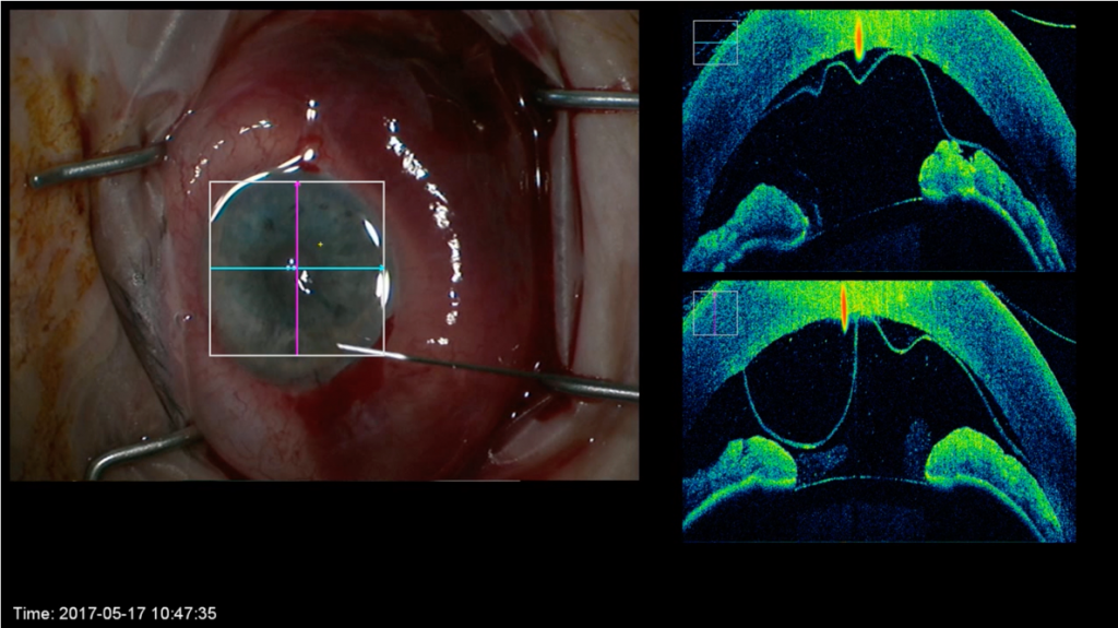 Intraoperative anterior segment OCT. Courtesy of Prof. Dr. Oliver Findl from Hanusch Hospital, Vienna, Austria.[/caption]
Does intraoperative anterior segment optical coherence tomography (iOCT) bring value to surgeons performing keratoplasty or just added cost?
Nino Hirnschall MD, PhD, and Massimo Busin MD presented their opposing views on this question in the Journal of Cataract & Refractive Surgery Symposium during the 36th Congress of the ESCRS in Vienna, Austria.
Dr Hirnschall, Hanusch Hospital Vienna, Austria, admitted that the technology has some limitations. Because a shadow is created behind metal instruments, transparent tools need to be developed for surgeons to take advantage of the full potential of OCT-guided imaging. In addition, with current technology, the iOCT scans are only two-dimensional. Nevertheless, there are benefits and evidence to support iOCT use during EK surgery, according to Dr Hirnschall.
“Intraoperative OCT may lead to safer surgery, and the only question is, if we need now, or in the future. But we definitely need it,” he said.
Discussing the advantages of iOCT, Dr Hirnschall said that because of the visualisation it provides, iOCT can allow DMEK in eyes with a cloudy cornea, where otherwise DSAEK would be needed by default. Because it clearly identifies Descemet remnants, iOCT also assures complete Descemet removal in DMEK and DSAEK.
“This is important because we know from several studies that the detachment rate is higher if the graft is placed on top of remnants of Descemet’s membrane,” Dr Hirnschall explained.
Evidence from published papers shows that by improving visualisation, iOCT in DMEK surgery can also be associated with shorter graft manipulation time that would be expected to translate into less endothelial cell loss. Furthermore, iOCT is useful in DMEK for confirming correct graft orientation.
“With iOCT, surgeons will never have an upside-down DMEK graft,” Dr Hirnschall said.
During both DMEK and DSAEK, iOCT enables identification of fluid in the donor-graft interface and of graft adherence. For deep anterior lamellar keratoplasty (DALK) or penetrating keratoplasty procedures, iOCT gives surgeons exact information about trephination depth and during DALK, it can guide cannula depth when creating the big bubble.
Prof Busin noted that results from the PIONEER and DISCOVER studies, in which surgeons indicated that iOCT affected decision-making in nearly one-half of cases, might be cited as evidence to support use of this technology. He pointed out, however, that the research has limitations.
“PIONEER and DISCOVER were not masked or randomised studies, and most importantly, they did not assess whether iOCT impacted actual clinical outcomes,” he said.
“The question we need to ask is whether the changed decision was relevant for the clinical outcome. We need more studies to demonstrate the true value of iOCT by showing that the clinical outcomes are better using the intraoperative imaging, rather than focusing on OCT-based outcomes, which may not be clinically relevant” said Prof Busin, Head, Department of Ophthalmology, Villa Igea Hospital, Forlì, Italy.
Prof Busin discussed a variety of techniques that can be used instead of iOCT to improve success rates in corneal lamellar transplant surgery. Although he said there is no evidence proving that residual Descemet’s membrane interferes with graft attachment, Prof Busin suggested that surgeons can visualise Descemet remnants by performing descemetorhexis under air.
He noted that surgeons can determine DMEK graft orientation by looking through the microscope. If they are unsure, there are a variety of effective, well-described strategies that are more practical and less expensive than iOCT, including a handheld slit beam, an S or F stamp, the Moutsouris sign or endoillumination.
Prof Busin also described surgical techniques he uses that obviate any need for iOCT for assessing interface fluid or cannula depth when injecting air for the big bubble.
To evacuate interface fluid in DSAEK cases, Prof Busin said he combines high-pressure tamponade with venting incisions. To achieve pneumatic dissection of the Descemet’s membrane in DALK, he precalibrates the trephine so that the blade advancement stops within 100 microns from the thinnest pachymetric value and the air cannula is inserted at the base of this trephination. In this technique, the depth of the cannula is based on predetermined quantitiative inputs and does require intraoperative clarification of depth from any source, including iOCT. Furthermore, in any case the cannulas currently available are metallic and therefore block the iOCT image in the most critical area; that is the residual stromal depth below the cannula.
Massimo Busin: massimo.busin@unife.it
Intraoperative anterior segment OCT. Courtesy of Prof. Dr. Oliver Findl from Hanusch Hospital, Vienna, Austria.[/caption]
Does intraoperative anterior segment optical coherence tomography (iOCT) bring value to surgeons performing keratoplasty or just added cost?
Nino Hirnschall MD, PhD, and Massimo Busin MD presented their opposing views on this question in the Journal of Cataract & Refractive Surgery Symposium during the 36th Congress of the ESCRS in Vienna, Austria.
Dr Hirnschall, Hanusch Hospital Vienna, Austria, admitted that the technology has some limitations. Because a shadow is created behind metal instruments, transparent tools need to be developed for surgeons to take advantage of the full potential of OCT-guided imaging. In addition, with current technology, the iOCT scans are only two-dimensional. Nevertheless, there are benefits and evidence to support iOCT use during EK surgery, according to Dr Hirnschall.
“Intraoperative OCT may lead to safer surgery, and the only question is, if we need now, or in the future. But we definitely need it,” he said.
Discussing the advantages of iOCT, Dr Hirnschall said that because of the visualisation it provides, iOCT can allow DMEK in eyes with a cloudy cornea, where otherwise DSAEK would be needed by default. Because it clearly identifies Descemet remnants, iOCT also assures complete Descemet removal in DMEK and DSAEK.
“This is important because we know from several studies that the detachment rate is higher if the graft is placed on top of remnants of Descemet’s membrane,” Dr Hirnschall explained.
Evidence from published papers shows that by improving visualisation, iOCT in DMEK surgery can also be associated with shorter graft manipulation time that would be expected to translate into less endothelial cell loss. Furthermore, iOCT is useful in DMEK for confirming correct graft orientation.
“With iOCT, surgeons will never have an upside-down DMEK graft,” Dr Hirnschall said.
During both DMEK and DSAEK, iOCT enables identification of fluid in the donor-graft interface and of graft adherence. For deep anterior lamellar keratoplasty (DALK) or penetrating keratoplasty procedures, iOCT gives surgeons exact information about trephination depth and during DALK, it can guide cannula depth when creating the big bubble.
Prof Busin noted that results from the PIONEER and DISCOVER studies, in which surgeons indicated that iOCT affected decision-making in nearly one-half of cases, might be cited as evidence to support use of this technology. He pointed out, however, that the research has limitations.
“PIONEER and DISCOVER were not masked or randomised studies, and most importantly, they did not assess whether iOCT impacted actual clinical outcomes,” he said.
“The question we need to ask is whether the changed decision was relevant for the clinical outcome. We need more studies to demonstrate the true value of iOCT by showing that the clinical outcomes are better using the intraoperative imaging, rather than focusing on OCT-based outcomes, which may not be clinically relevant” said Prof Busin, Head, Department of Ophthalmology, Villa Igea Hospital, Forlì, Italy.
Prof Busin discussed a variety of techniques that can be used instead of iOCT to improve success rates in corneal lamellar transplant surgery. Although he said there is no evidence proving that residual Descemet’s membrane interferes with graft attachment, Prof Busin suggested that surgeons can visualise Descemet remnants by performing descemetorhexis under air.
He noted that surgeons can determine DMEK graft orientation by looking through the microscope. If they are unsure, there are a variety of effective, well-described strategies that are more practical and less expensive than iOCT, including a handheld slit beam, an S or F stamp, the Moutsouris sign or endoillumination.
Prof Busin also described surgical techniques he uses that obviate any need for iOCT for assessing interface fluid or cannula depth when injecting air for the big bubble.
To evacuate interface fluid in DSAEK cases, Prof Busin said he combines high-pressure tamponade with venting incisions. To achieve pneumatic dissection of the Descemet’s membrane in DALK, he precalibrates the trephine so that the blade advancement stops within 100 microns from the thinnest pachymetric value and the air cannula is inserted at the base of this trephination. In this technique, the depth of the cannula is based on predetermined quantitiative inputs and does require intraoperative clarification of depth from any source, including iOCT. Furthermore, in any case the cannulas currently available are metallic and therefore block the iOCT image in the most critical area; that is the residual stromal depth below the cannula.
Massimo Busin: massimo.busin@unife.it