Image-Guided Toric IOL
New technology makes axis alignment easier for better outcomes
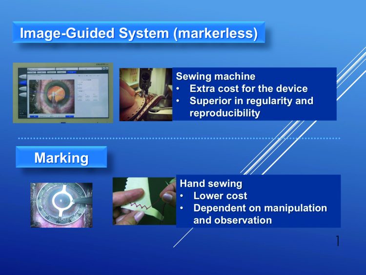 The difference between the image-guided system and conventional marking. Courtesy of Hiroko Bissen-Miyajima MD, PhD
In just six years, new technology has revolutionised implantation of toric intraocular lenses (IOLs), Hiroko Bissen-Miyajima MD, PhD told the 2016 ASCRS•ASOA Symposium & Congress in New Orleans, USA. Image-guided systems are doing away with manually marking the astigmatism axis, greatly increasing lens alignment precision.
When Dr Bissen-Miyajima, who is Professor and Chair of the Ophthalmology Department at Tokyo Dental College, Japan, wrote her Japanese language text on toric IOLs in 2010, experts from around the world shared techniques for manually marking the astigmatism axis on the cornea and conjunctiva before surgery. They then confirmed the axis by superimposing it on a topography map. During surgery the compressed mark remained as a reference for manually aligning the lens, she said. Yet even the width of the mark can take up three to five degrees, limiting its precision.
In 2016, image-guided systems, such as Carl Zeiss Meditec’s CALLISTO eye and Alcon’s VERION, make the process much easier, Dr Bissen-Miyajima said. During diagnostic workup, a reference image is taken of the eye and the axis noted. During surgery, the device matches the blood vessel landmarks seen through the microscope with those of the reference image, and projects the axis in the eyepiece as a reference for aligning the IOL.
SEWING MACHINE
Dr Bissen-Miyajima likened the markerless image-guided system to a sewing machine. By comparison, manual marking is more like hand sewing. “There is extra cost for the device but it is superior in regularity and reproducibility. While it is lower in cost, manual marking is dependent on manipulation and observation by the surgeon,” she said.
Other new technologies will further improve the precision and outcomes of toric IOLs, Dr Bissen-Miyajima said. These include measuring anterior and posterior corneal astigmatism, which will allow more accurate toric power calculations and more precise reference axis. Digital analysis of IOL position, including axis alignment, will enable precise alignment corrections during surgery. Intraoperative aberrometry will help determine the proper axis after lens removal. In addition, image-guided femtosecond lasers will better control incisions, astigmatic correction and induced astigmatism.
Dr Bissen-Miyajima was one of the ASCRS’ Honoured Guests at this year’s symposium. She was lauded for her work investigating multifocal IOLs and femtosecond laser-assisted cataract surgery, her work as a reviewer for several journals worldwide, as President of the Japanese Society of Cataract and Refractive Surgery, and for her leadership in several international societies.
Hiroko Bissen-Miyajima: bissen@tdc.ac.jp
Published
Wednesday, January 10, 2018
Category