DSAEK continues to serve a purpose in corneal transplantation
Many complex cases are better suited to UT-DSAEK

Dermot McGrath
Published: Saturday, September 14, 2019
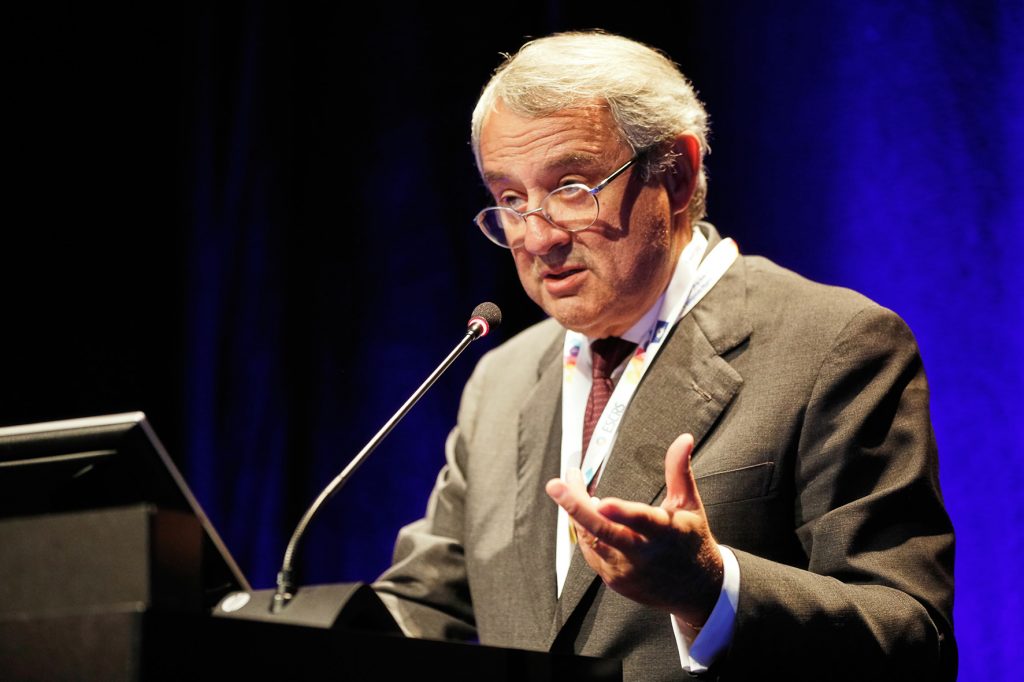 Massimo Busin[/caption]
ALTHOUGH corneal transplantation surgery has seen an increase in Descemet Membrane Endothelial Keratoplasty (DMEK) in recent years for the reported benefits of quicker healing and better visual outcomes, procedures such as ultrathin Descemet Stripping Automated Endothelial Keratoplasty (UT-DSAEK) still have a valuable role to play for certain indications, according to Massimo Busin MD.
“DMEK is certainly a viable option for indications such as Fuchs’ endothelial dystrophy and gives excellent results, but there are still many complex cases, with poor visibility and certain comorbidities for instance, that are better suited to UT-DSAEK,” he told delegates attending the 10th EuCornea Congress in Paris.
Issues to bear in mind when considering UT-DSAEK versus DMEK is the preoperative eye status, surgical technique, outcomes and complications, noted Dr Busin. In his view, both procedures have a role to play depending on the needs of each individual patient.
DMEK is a good option, for instance in patients with Fuchs’ endothelial dystrophy, intact posterior capsule and normal anterior segment anatomy. However, UT-DSAEK is better adapted to cases with a shallow or poorly visualised anterior chamber or more complex eyes with ocular comorbidities such as glaucoma or severe pseudophakic bullous keratopathy, and aphakic or vitrectomised eyes.
Massimo Busin[/caption]
ALTHOUGH corneal transplantation surgery has seen an increase in Descemet Membrane Endothelial Keratoplasty (DMEK) in recent years for the reported benefits of quicker healing and better visual outcomes, procedures such as ultrathin Descemet Stripping Automated Endothelial Keratoplasty (UT-DSAEK) still have a valuable role to play for certain indications, according to Massimo Busin MD.
“DMEK is certainly a viable option for indications such as Fuchs’ endothelial dystrophy and gives excellent results, but there are still many complex cases, with poor visibility and certain comorbidities for instance, that are better suited to UT-DSAEK,” he told delegates attending the 10th EuCornea Congress in Paris.
Issues to bear in mind when considering UT-DSAEK versus DMEK is the preoperative eye status, surgical technique, outcomes and complications, noted Dr Busin. In his view, both procedures have a role to play depending on the needs of each individual patient.
DMEK is a good option, for instance in patients with Fuchs’ endothelial dystrophy, intact posterior capsule and normal anterior segment anatomy. However, UT-DSAEK is better adapted to cases with a shallow or poorly visualised anterior chamber or more complex eyes with ocular comorbidities such as glaucoma or severe pseudophakic bullous keratopathy, and aphakic or vitrectomised eyes.
Tags: dmek, Ultrathin DSAEK
Latest Articles
Towards a Unified IOL Classification
The new IOL functional classification needs a strong and unified effort from surgeons, societies, and industry.
Organising for Success
Professional and personal goals drive practice ownership and operational choices.
Update on Astigmatism Analysis
Is Frugal Innovation Possible in Ophthalmology?
Improving access through financially and environmentally sustainable innovation.
iNovation Innovators Den Boosts Eye Care Pioneers
New ideas and industry, colleague, and funding contacts among the benefits.
José Güell: Trends in Cornea Treatment
Endothelial damage, cellular treatments, human tissue, and infections are key concerns on the horizon.
Making IOLs a More Personal Choice
Surgeons may prefer some IOLs for their patients, but what about for themselves?
Need to Know: Higher-Order Aberrations and Polynomials
This first instalment in a tutorial series will discuss more on the measurement and clinical implications of HOAs.
Never Go In Blind
Novel ophthalmic block simulator promises higher rates of confidence and competence in trainees.