Coats’ disease
New classification for the rare condition proposed

Dermot McGrath
Published: Friday, February 1, 2019
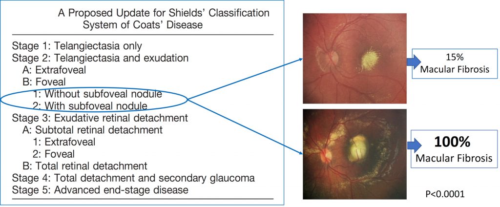 Proposed update for Shields classification of Coats disease. Two subcategories have been introduced within Stage 2B, without or with subfoveal nodule (blue circle). The presence of a subfoveal nodule means a higher risk of macular fibrosis and poor visual outcome compared with patients with flat exudation (100% vs 15%, p<0.0001)
The onset of Coats’ disease in children of a younger age is associated with more severe manifestations, more advanced disease stage at diagnosis and worse visual outcome. Age, correlated with disease stage, should be considered a prognostic marker in Coats’ disease, according to Alejandra Daruich, MD (Daruich A et al. Retina 2018).
“Coats’ disease is a rare condition, affecting about one in every 100,000 children, it is usually unilaterally and affects young boys with a mean age of 5 or 6 years. In children younger than 4 years old leukocoria or strabismus are the main manifestations, but older children could be asymptomatic so we need to be particularly vigilant with these patients,” she told delegates attending the World Society of Paediatric Ophthalmology and Strabismus (WSPOS) subspecialty day at the 36th Congress of the ESCRS in Vienna.
Peripheral telangiectasias of the retinal vasculature are the main manifestation in Coats’ disease, noted Dr Daruich, adding that the telangiectatic vessels are rarely located in the macula. These telangiectasias consist of dilated capillaries that provoke intraretinal and subretinal exudation. This process may eventually lead to exudative retinal detachment, neovascular glaucoma and profound vision loss.
Intraretinal and subretinal exudates often affect areas of the retina remote from the telangiectasias and tend to migrate toward the macula, although there is no clear explanation as to why the exudation preferentially accumulates in the macula, she said.
The most commonly used classification system for Coats’ disease derives from work by Shields et al. and takes into account the presence and location of lipid exudates at presentation to stratify the visual prognosis of the disease.
“We have proposed updating Shields’ classification of Coats’ disease to include two subcategories within Stage 2B relating to foveal exudation. These categories would be with or without subfoveal nodule as we have shown (Daruich A et al. Retina. 2017) that the presence of a subfoveal nodule means a higher risk of macular fibrosis and poor visual outcome for these patients,” she said.
Although Coats’ has long been considered an unilateral disease, some recent studies using optical coherence tomography angiography (OCT-A) and fluorescein angiography (FA) indicate contralateral abnormal peripheral vasculature in some patients.
“The presence of bilateral asymmetric disease may be higher than previously thought although this needs further investigation,” she said.
Treatment for Coats’ disease is usually laser photocoagulation to the telangiectasias or cryotherapy depending on the stage of the disease. Although anti-VEGF has been used as adjuvant therapy in advanced cases of Coats’ disease, its use may carry a higher risk of vitreoretinal fibrosis and tractional retinal detachment.
“We need more data to be able to support anti-VEGF treatment as an adjunctive therapy in these young patients,” she concluded.
Alejandra Daruich: adaruich.matet@gmail.com
Proposed update for Shields classification of Coats disease. Two subcategories have been introduced within Stage 2B, without or with subfoveal nodule (blue circle). The presence of a subfoveal nodule means a higher risk of macular fibrosis and poor visual outcome compared with patients with flat exudation (100% vs 15%, p<0.0001)
The onset of Coats’ disease in children of a younger age is associated with more severe manifestations, more advanced disease stage at diagnosis and worse visual outcome. Age, correlated with disease stage, should be considered a prognostic marker in Coats’ disease, according to Alejandra Daruich, MD (Daruich A et al. Retina 2018).
“Coats’ disease is a rare condition, affecting about one in every 100,000 children, it is usually unilaterally and affects young boys with a mean age of 5 or 6 years. In children younger than 4 years old leukocoria or strabismus are the main manifestations, but older children could be asymptomatic so we need to be particularly vigilant with these patients,” she told delegates attending the World Society of Paediatric Ophthalmology and Strabismus (WSPOS) subspecialty day at the 36th Congress of the ESCRS in Vienna.
Peripheral telangiectasias of the retinal vasculature are the main manifestation in Coats’ disease, noted Dr Daruich, adding that the telangiectatic vessels are rarely located in the macula. These telangiectasias consist of dilated capillaries that provoke intraretinal and subretinal exudation. This process may eventually lead to exudative retinal detachment, neovascular glaucoma and profound vision loss.
Intraretinal and subretinal exudates often affect areas of the retina remote from the telangiectasias and tend to migrate toward the macula, although there is no clear explanation as to why the exudation preferentially accumulates in the macula, she said.
The most commonly used classification system for Coats’ disease derives from work by Shields et al. and takes into account the presence and location of lipid exudates at presentation to stratify the visual prognosis of the disease.
“We have proposed updating Shields’ classification of Coats’ disease to include two subcategories within Stage 2B relating to foveal exudation. These categories would be with or without subfoveal nodule as we have shown (Daruich A et al. Retina. 2017) that the presence of a subfoveal nodule means a higher risk of macular fibrosis and poor visual outcome for these patients,” she said.
Although Coats’ has long been considered an unilateral disease, some recent studies using optical coherence tomography angiography (OCT-A) and fluorescein angiography (FA) indicate contralateral abnormal peripheral vasculature in some patients.
“The presence of bilateral asymmetric disease may be higher than previously thought although this needs further investigation,” she said.
Treatment for Coats’ disease is usually laser photocoagulation to the telangiectasias or cryotherapy depending on the stage of the disease. Although anti-VEGF has been used as adjuvant therapy in advanced cases of Coats’ disease, its use may carry a higher risk of vitreoretinal fibrosis and tractional retinal detachment.
“We need more data to be able to support anti-VEGF treatment as an adjunctive therapy in these young patients,” she concluded.
Alejandra Daruich: adaruich.matet@gmail.com