Anaridia-associated keratopathy
Research revealing genetic clues to aniridia-associated keratopathy.

Roibeard O’hEineachain
Published: Friday, November 1, 2019
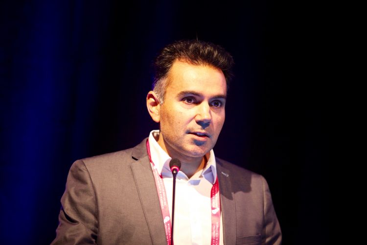
 Neil Lagali PhD speaking at the WSPOS sub-specialty day in Paris
Research is continuing to reveal the multifaceted nature of aniridia-associated keratopathy (AAK), said Associate Professor Neil Lagali PhD, Institution for Clinical and Experimental Medicine, Linköping University, Sweden, at the World Society of Paediatric Ophthalmology and Strabismus (WSPOS) sub-specialty day in Paris, France.
He noted that congenital aniridia is a rare but serious inherited eye disease requiring a lifetime of ophthalmic care, starting in infancy. The condition has a prevalence of one per 100,000 population. It is primarily caused by mutations in the PAX6 gene coding for the PAX6 protein. More than 400 such mutations in the PAX6 gene have thus far been identified.
The defining characteristic of congenital aniridia is a partial or total iris hypoplasia. The phenotype also often involves aberrant development of the eye’s drainage channels, leading to glaucoma. Patients also tend to develop early cataract and most have nystagmus, as well as an underdeveloped fovea. In addition, congenital aniridia patients usually have limbal stem cell insufficiency, which, over time, leads to aniridia-associated keratopathy (AAK).
Prof Lagali noted that although the pathobiology of AAK is not completely understood, it may result from a breakdown in the limbal stem cells leading to conjunctivalisation of the entire corneal surface. AAK is difficult to treat with keratoplasty, because of the associated high risk of graft rejection and inadequate wound closure. Keratoplasty in these patients also carries the risk of aniridia fibrosis syndrome, a condition that results in fibrotic structures developing in the anterior chamber.
However, AAK is more than limbal stem cell deficiency, he noted. Other contributing factors include meibomian gland dysfunction and loss of meibomian gland cells, reduced tear production and increased tear film osmolarity. In addition, inflammatory mediators play a role.
Prof Lagali reported a study in which he and his associates in Norway, led by Prof Tor Utheim, obtained tear samples of 35 persons with aniridia and 21 controls. The study showed up-regulation of six different immunomodulators, but also showed reduced levels of the IL-1β-antagonist, IL1-RA.
“Tear film activation of these immunomodulators could stimulate inflammatory cells to invade the cornea that can lead to rejection of any kind of implanted tissue,” Prof Lagali said.
He added that genotype testing is critical for prognosis of AAK. In another recently published study, he and his colleagues in Germany, including Prof Barbara Käsmann-Kellner and Prof Berthold Seitz, found that around 70% of AAK cases have PTC/CTE mutations, which cause a progressive form of the disease, leading to eventual conjunctivalisation of the entire cornea. Some 10% have entire chromosomal deletions, which lead to an aggressive phenotype at a young age.
On the other hand, around 10% of patients have PAX6 non-coding mutations, which result in a mild, non-progressive phenotype, as is similarly the case with the 10% having missense mutations.
Moving forward, Prof Lagali noted that patient stratification by genotype is important not only for prognosis but for evaluating the effect of potential treatments for AAK in the future.
Neil Lagali: neil.lagali@liu.se
Acknowledgment: The work described here was in part supported by the European Union’s COST Program, under COST Action CA18116, ANIRIDIA-NET
(www.aniridia-net.eu)
Tags: anaridia associated keratopathy
Latest Articles
Organising for Success
Professional and personal goals drive practice ownership and operational choices.
Update on Astigmatism Analysis
Is Frugal Innovation Possible in Ophthalmology?
Improving access through financially and environmentally sustainable innovation.
iNovation Innovators Den Boosts Eye Care Pioneers
New ideas and industry, colleague, and funding contacts among the benefits.
Making IOLs a More Personal Choice
Surgeons may prefer some IOLs for their patients, but what about for themselves?
Need to Know: Higher-Order Aberrations and Polynomials
This first instalment in a tutorial series will discuss more on the measurement and clinical implications of HOAs.
Never Go In Blind
Novel ophthalmic block simulator promises higher rates of confidence and competence in trainees.
Simulators Benefit Surgeons and Patients
Helping young surgeons build confidence and expertise.
How Many Surgeries Equal Surgical Proficiency?
Internet, labs, simulators, and assisting surgery all contribute.